Understanding Sciatica: Causes, Treatment Options, and Prognosis
Introduction:
Sciatica is an often debilitating condition that typically appears after age 60 in those affected, characterized by dysthesias: abnormal sensations that can include shooting pain, numbness and tingling radiating (traveling) along the path of the sciatic nerve, the largest diameter nerve in the body, which runs from the lower back, between the deep hip rotator muscles, and down the back of each leg. This condition can significantly impair one’s quality of life, affecting mobility, work, and daily activities. In this post, I’ll delve into the main details of sciatica, exploring its causes, pathology, treatment options ranging from conservative approaches to surgical interventions, and the prognosis associated with each.
Understanding the Pathology of Sciatica:
Sciatica typically arises from compression or irritation of the sciatic nerve roots, also called the cauda equina, most commonly at the lumbar spine level. The sciatic nerve is composed of nerve roots originating from the lumbar and sacral spine (L4-S3). When these nerve roots are compressed or inflamed, they can give rise to the characteristic symptoms of sciatica, including pain, numbness, tingling, and weakness along the nerve’s distribution. The sciatic nerve is comprised of both motor and sensory fibers, but since the sensory fibers are larger in diameter they are more susceptible to mechanical pressure; hence, irritation of the nerves results in mostly sensory dysfunction and less of motor function (leg muscle strength and coordination).
Common Causes of Sciatica
- Herniated Disc: One of the leading causes of sciatica is a herniated disc, also known as a slipped or ruptured disc. Intervertebral discs act as cushions between the vertebrae of the spine, providing support and flexibility. When a disc herniates, its inner gel-like material protrudes through the tough outer layer, exerting pressure on nearby nerve roots, including those of the sciatic nerve.
- Spinal Stenosis: Spinal stenosis refers to the narrowing of the spinal canal: the passageway formed from the stacking of the spinal vertebrae, which are solid in the front and have a ringed rear portion that when stacked form the canal in which the spinal cord resides. Narrowing can occur due to age-related degenerative changes, such as the formation of bone spurs and thickening of ligaments. The bone spurs and buckled ligaments encroach the canal, narrowing it. This narrowing can compress the spinal cord and nerve roots, or cause them to rub against them during movements especially back extension, leading to sciatic symptoms.
- Piriformis Syndrome: The piriformis muscle, located in the buttocks region, plays a crucial role in hip rotation. In some individuals, the sciatic nerve may pass through or under the piriformis muscle, making it susceptible to compression or irritation. This condition, known as piriformis syndrome, can mimic the symptoms of sciatica. The muscles scissor the nerve if they get spasmed, which can produce sciatica symptoms.
- Spondylolisthesis: Spondylolisthesis occurs when a vertebra slips out of alignment anteriorly, often due to degenerative changes or trauma (fractured pars). This misalignment offsets the foramen at that level, usually at L4’L5 effectively scissoring the nerve roots and producing sciatica symptoms.
- Degenerative Disc Disease: With age, the intervertebral discs undergo wear and tear, leading to degenerative changes such as disc dehydration, loss of disc height, and the formation of bone spurs. These changes can contribute to nerve root compression and the development of sciatica.
Treatment Options for Sciatica:
The management of sciatica aims to alleviate pain, reduce inflammation, improve mobility, and address the underlying cause of the condition. Treatment options may vary depending on the severity of symptoms, the underlying pathology, and individual patient factors.
- Conservative Management: Conservative approaches are often the first line of treatment for sciatica and may include:
- Pain Medications: Nonsteroidal anti-inflammatory drugs (NSAIDs), muscle relaxants, and analgesics can help alleviate pain and reduce inflammation.
- Physical Therapy: Targeted exercises, stretches, and manual techniques can improve spinal flexibility, strengthen supporting muscles, and alleviate pressure on the sciatic nerve.
- Heat and Cold Therapy: Applying heat or cold packs to the affected area can help reduce pain and inflammation.
- Epidural Steroid Injections: Corticosteroids injected into the epidural space can provide temporary relief by reducing inflammation around the affected nerve roots.
- Surgical Intervention: If conservative measures fail to provide adequate relief or if there is evidence of progressive neurological deficit, surgical intervention may be considered. Surgical options for sciatica include:
- Discectomy: In cases of herniated discs causing nerve compression, a discectomy may be performed to remove the protruding disc material and relieve pressure on the affected nerve roots.
- Laminectomy: This procedure involves removing a portion of the vertebral bone (lamina) to alleviate pressure on the spinal cord and nerve roots, particularly in cases of spinal stenosis.
- Spinal Fusion: Spinal fusion surgery may be recommended to stabilize the spine and prevent further slippage of vertebrae in cases of spondylolisthesis or severe degenerative disc disease.
- Alternative Therapies: Some individuals may find relief from sciatica symptoms through alternative therapies, although evidence supporting their efficacy may vary. These may include:
- Acupuncture: The insertion of fine needles into specific points on the body may help reduce pain and improve nerve function.
- Chiropractic Care: Spinal manipulation techniques performed by trained chiropractors may help alleviate pressure on the sciatic nerve and improve spinal alignment. Combining chiropractic with a stretching and exercise routine is even better.
- Mechanical Traction: Some chiropractic and physical therapy clinics have special tables that can stretch the spine using an electric motor. This may increase space between the vertebrae, retract buckled ligaments and provide temporary relief.
- Yoga and Pilates: These forms of exercise focus on strengthening core muscles, improving flexibility, and promoting relaxation, which can be beneficial for individuals with sciatica.
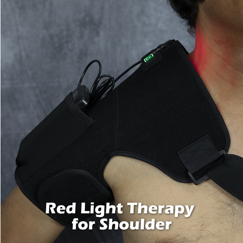
- Low Level Laser (LLLT): Lasering the area of the sciatic nerve may alleviate symptoms. LLLT, also known as cold laser (non-thermal) helps by providing deep penetrating light to the nerve tissue. Photons from laser light enter the sciatic nerve and can modulate pain producing biochemical pathways.
Prognosis:
The prognosis for sciatica depends on various factors, including the underlying cause, the severity of symptoms, and the effectiveness of treatment. In many cases, sciatica resolves with conservative measures within a few weeks to months. However, some individuals may experience chronic or recurrent symptoms that require ongoing management. Over time, the neurons in the irritated nerve roots lose some of their ability to conduct sensory signals, and the symptoms tend to be less acute.
Surgical intervention can provide significant relief for those with severe or persistent symptoms, but it also carries risks and requires careful consideration of potential benefits versus potential complications. With advances in surgical techniques and rehabilitation protocols, the outcomes of surgical treatment for sciatica have improved, with many patients experiencing long-term symptom relief and improved function.
Ultimately, the prognosis for sciatica is influenced by factors such as the individual’s overall health, adherence to treatment recommendations, and the presence of any underlying medical conditions. Early intervention, comprehensive management strategies, and a multidisciplinary approach involving healthcare providers from various specialties can optimize outcomes and improve the quality of life for individuals affected by sciatica.
Conclusion:
Sciatica is a complex condition with diverse causes, ranging from herniated discs to spinal stenosis and piriformis syndrome. Understanding the underlying pathology is crucial for guiding appropriate treatment interventions, which may include conservative measures, surgical intervention, and alternative therapies. With timely and comprehensive management, the prognosis for sciatica can be favorable, enabling individuals to regain function and resume their daily activities with minimal pain and discomfort.


 foramen (canal), or space where the spinal cord resides. When there is less space for the spinal cord to move, it is subject to more abrasion with spinal movement; i.e. bending and turning your neck. The cord (actually, meninges or covering of the cord) rubs against sharp edges of the bony projections into the foramen with movement causing inflammation and injury to the nerve tissue, sometimes causing sclerosis (hardening). In advanced cases, especially cases of lumbar spinal stenosis (due to the more significant weight burden) the narrowing gets so advanced that there is constant pressure on the nerve roots. At this point, it is an emergency situation as renal function and sensation to the legs are affected.